


Gynecomastia Surgery for Puffy Nipples in a 27-Year-Old
A 27-year-old professional from Hyderabad presented with bilateral puffy nipples and visible chest enlargement that had been present since his late teens. Clinically assessed as Grade I gynecomastia with both a glandular and a fatty component, the condition had not responded to any conservative measure and was causing significant self-consciousness in daily and professional settings. Dr. Harikiran Chekuri at Redefine Clinic performed liposuction combined with direct gland excision under general anaesthesia. The procedure was completed in 40 minutes, the patient recovered from anaesthesia immediately, and the chest was flat by the first follow-up at day three.
PATIENT PROFILE
| PARAMETER | DETAIL |
| Age | 27 years |
| Gender | Male |
| City | Hyderabad, India |
| Presenting Complaint | Bilateral puffy nipples, visible chest enlargement, self-consciousness in fitted clothing and social settings |
| Diagnosis | Grade I Gynecomastia (mixed glandular and fatty component, bilateral) |
| BMI | Within normal range |
| Duration of Condition | Present since late teens. No spontaneous resolution. |
| Hormonal Workup | Conducted and reviewed prior to surgery. No pathological cause identified. |
| Anaesthetic Assessment | Completed by anaesthetist. Cleared for general anaesthesia. |
| Previous Treatments | None surgical. Conservative measures attempted without improvement. |
| Drug or Supplement History | None relevant disclosed |
| Date of Surgery | July 2026 |
| Outcome | Excellent |
THE PROBLEM
The puffy nipples had been there as long as he could remember. Through his teens and into his mid-twenties, he had assumed it was a phase that would resolve. It did not. The chest had a visible fullness that no amount of weight management or exercise had shifted. The nipples sat proud and rounded under any fitted clothing, making him self-conscious in situations most people never think about. Gym changing rooms, beach outings, fitted shirts for work. Each one required a level of mental effort it should not have taken.
He had no history of steroid use, no medication that could account for the finding, and a hormonal workup that came back entirely within normal range. The cause was idiopathic. The effect was real. He had been managing it with clothing choices for almost a decade and had reached the point where he wanted a permanent answer rather than another workaround.
After researching gynecomastia surgery in Hyderabad he came in for a consultation at Redefine. He wanted to understand exactly what the surgery involved, how long the recovery would take, and what the result would look like.
CONSULTATION & TREATMENT PLAN
WHAT WAS ASSESSED DURING THE CONSULTATION
A complete clinical and investigative assessment was conducted before any surgical recommendation was made:
-
- Physical examination: bilateral Grade I gynecomastia confirmed. Firm glandular disc palpable beneath each nipple-areola complex. Moderate periareolar fatty component present on both sides contributing to the puffy, rounded nipple appearance.
- Grade assessment: Grade I confirmed bilaterally. Glandular tissue present with associated periareolar fat. No skin excess. No ptosis.
- Hormonal workup reviewed: testosterone, LH, FSH, estradiol, prolactin, and thyroid function all within normal range. No pathological cause identified.
- Anaesthetist assessment: patient reviewed and cleared for general anaesthesia prior to scheduling.
- Surgical markings documented: midline reference, periareolar gland excision zones, and liposuction zones marked bilaterally with cross-hatching to guide fat removal above and around each breast mound.
- Patient goals confirmed: flat chest, natural contour, no visible scarring, quick return to normal activity.
- Follow-up schedule discussed: day 3 review, one-week review, and ongoing compression garment compliance confirmed.
WHY LIPOSUCTION WITH GLAND EXCISION WAS CHOSEN
-
- Grade I gynecomastia with both a glandular and fatty component requires a two-part approach. Liposuction alone cannot remove the firm central gland. Gland excision alone would leave the periareolar fat intact and result in an incomplete contour improvement.
- Liposuction of the periareolar fat zone was performed first to reduce bulk, improve access to the gland, and allow the skin to retract naturally over the flattened chest.
- Direct gland excision through a minimal periareolar incision then removed the firm glandular disc completely, addressing the root cause of the nipple puffiness.
- Combined approach in a single 40-minute session avoids the need for staged procedures and achieves both contour improvement and gland removal simultaneously.
- General anaesthesia selected for patient comfort and to allow a completely still operative field for precise excision. Immediate recovery from anaesthesia was noted and the patient was discharged the same day.
- No drains required given the limited scope and short operative time.
PRE-OPERATIVE PHOTOS
Pre-operative photographs and surgical markings were documented from frontal and bilateral oblique angles. Markings show midline reference, bilateral periareolar excision zones, and cross-hatched liposuction zones. Intraoperative photographs confirm markings under theatre conditions.
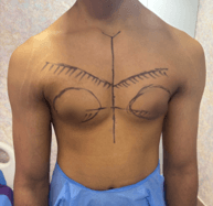
Pre-op: frontal view
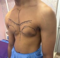
Pre-op: right oblique
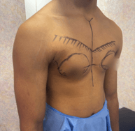
Pre-op: left oblique
PROCEDURE DETAILS
- Patient positioned supine under general anaesthesia. Surgical markings confirmed and reviewed by Dr. Harikiran Chekuri before incision.
- Tumescent solution infiltrated bilaterally into the periareolar fat zones and the cross-hatched liposuction areas to reduce bleeding and facilitate fat aspiration.
- Liposuction performed through 3mm access ports at the lateral chest border bilaterally, targeting the periareolar fat zones marked pre-operatively.
- Periareolar incision made at the inferior areolar border on the right side. Glandular disc dissected and excised completely as a single specimen.
- Haemostasis achieved on the right. Procedure repeated identically on the left side.
- Complete gland removal confirmed by palpation on both sides. No residual firmness detected.
- Wounds closed in layers with absorbable deep sutures and fine subcuticular absorbable suture at skin level within the periareolar border on each side.
- Compression garment applied in theatre immediately following wound closure.
- Total operative time 40 minutes. Patient recovered from general anaesthesia immediately. Discharged same day.
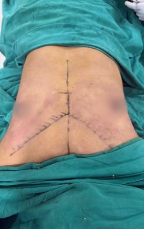
Intraoperative: supine, markings (frontal)
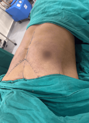
Intraoperative: supine, markings (oblique)
PROCEDURE FACTS
| PARAMETER | DETAIL |
| Procedure | Liposuction and Direct Gland Excision (Bilateral) |
| Duration | 40 minutes |
| Anaesthesia | General anaesthesia |
| Liposuction | Periareolar fat zones, bilateral, via 3mm lateral port access |
| Gland Excision | Complete bilateral glandular disc excision via periareolar incision |
| Incision | Inferior periareolar border, bilateral. Scar within areolar border. |
| Drains | Not required |
| Anaesthesia Recovery | Immediate |
| Hospital Stay | Day care. Discharged same day. |
| Complications | None |
POST-OPERATIVE RESULTS
The result was evident at the first follow-up on day three. Both nipples lay flat against the chest wall with no residual puffiness. Mild swelling was present but the improvement in chest contour was already clear. At the one-week review, swelling had reduced further and the periareolar wounds were healing cleanly within the areolar border. The patient reported no significant discomfort beyond the first two days and had returned to light daily activity by day four. Compression garment compliance was confirmed at both early reviews. At the six-week mark, the chest was flat and symmetric, the periareolar scars were imperceptible within the areolar skin texture, and the patient confirmed the result had fully met his expectations.
OUTCOMES AT A GLANCE
| OUTCOME METRIC | RESULT |
| Chest Contour | Flat and symmetric bilaterally from day 3 review onwards |
| Nipple Appearance | No residual puffiness. Nipples lying flat against chest wall. |
| Glandular Tissue | Completely excised bilaterally. Confirmed intraoperatively. |
| Periareolar Fat | Reduced by liposuction. Smooth contour transition achieved. |
| Scar Visibility | Imperceptible at 6 weeks. Scar within areolar border on both sides. |
| Symmetry | Excellent bilateral symmetry |
| Return to Activity | Light daily activity from day 4. Normal activity from week 2. |
| Compression Garment | Worn continuously for 6 weeks as instructed |
| Complications | None |
| Outcome | Excellent |
PATIENT FEEDBACK
Feedback recorded at the 6-week follow-up visit at Redefine, Hyderabad.
Google Review
★ ★ ★ ★ ★ 5.0
Verified Patient (Name withheld for privacy)
“I had been dealing with this since I was a teenager. Almost ten years of choosing the right shirts so no one would notice. When I finally came to Redefine, Dr. Harikiran explained the procedure clearly and told me the surgery would take around 40 minutes. I was not expecting such a short operative time. I woke up from anaesthesia immediately and was home the same evening. By day three when I came for my first follow-up, the difference was already visible. Six weeks in, the chest is flat, the scars are invisible, and I have stopped thinking about what I am wearing for the first time in years. I should have done this much sooner.”
Profile: Male, 27 years, Hyderabad
Procedure: Liposuction and Gland Excision (Grade I Gynecomastia), Redefine Gachibowli, July 2026
Doctor: Dr. Harikiran Chekuri, Redefine
Note: Due to privacy regulations, the patient’s name is not displayed. This review has been shared with the patient’s written consent.
POST-OPERATIVE CARE & RECOVERY
- Wear compression garment continuously for 6 weeks, preferably 24 hours a day as instructed at discharge.
- Take prescribed analgesics for discomfort in the first 3 days as directed. Do not exceed the stated dose.
- Apply antiseptic ointment to the periareolar incision sites as instructed until the one-week review.
- Take prescribed multivitamin daily throughout the recovery period to support healing.
- Maintain a high protein diet throughout the recovery period. Adequate protein supports wound healing and tissue repair.
- Keep the surgical sites dry for 7 days. No bathing, swimming, or direct water contact on the incisions before the one-week review.
- No lifting, pushing, or upper body exercise for 4 weeks.
- Light walking permitted from day 2 onwards. Resume normal daily activity from week 2.
- First follow-up at day 3. Second follow-up at one week. Further reviews as advised.
RECOVERY TIMELINE
| TIMEFRAME | WHAT TO EXPECT |
| Day 1 to 2 | Mild discomfort and chest tightness. Compression garment worn. Rest at home. Prescribed analgesics taken. |
| Day 3 | First follow-up. Wound check. Swelling present but chest contour clearly improved. Continue antiseptic ointment and compression. |
| Day 4 to 7 | Discomfort resolving. Light daily activity resumed. Second follow-up at one week for wound review and dressing change. |
| Week 2 to 4 | Swelling reducing. Chest contour improving progressively. Compression garment continues 24 hours a day. Normal daily activity from week 2. |
| Week 4 to 6 | Final weeks of compression garment. Periareolar scars fading within areolar border. Upper body exercise avoided until week 6 clearance. |
| Week 6 onwards | Compression garment discontinued. Full activity resumed. Final result confirmed. Scars continuing to fade. |
DISCLAIMER:
This case study is for informational purposes only and does not constitute medical advice. Individual results may vary based on the grade of gynecomastia, body composition, skin elasticity, and individual healing response. A thorough hormonal workup and anaesthetic assessment are required before any gynecomastia surgery is undertaken. Consult a qualified plastic surgeon to assess your specific condition and discuss what outcome is realistic for your presentation. Patient identity withheld per confidentiality guidelines. Feedback published with written consent.